
(2)Columnaris
From Wikipedia, the free encyclopedia
Jump to: navigation, search
Columnaris in a Chinook salmon
is a symptom of disease in fish which results from an infection caused by the bacteria
Flavobacterium columnare. The disease is highly contagious and the outcome is often fatal.
Causes
The bacterium usually enters fish through gills, mouth, or small wounds, and is prevalent where high bio-load exists, or where conditions may be stressful due to overcrowding or low dissolved oxygen levels in the water column. The bacteria can persist in water for up to 32 days when the hardness is 50ppm or more
[1].
Symptoms
An infection will usually first manifest in fish by causing frayed and ragged fins. This is followed by the appearance of ulcerations on the skin, and subsequent epidermal loss, identifiable as white or cloudy fungus-like patches. Gills will change colour, either becoming light or dark brown, and may also manifest necrosis. Fish will breathe rapidly and laboriously as a sign of gill damage.
Prognosis
Ulcerations develop within 24 to 48 hours. Fatality occurs between 48 to 72 hours if no treatment is pursued; however, at higher temperatures death may occur within hours.
Treatment
Medicated food containing oxytetracycline is an effective treatment. Sulfate based drug combinations such as TMP Sulfa, Sulfa 4 TMP, or Triple Sulfa also combat the infection. Erythromycin (Maracyn), nitrofurazone, nifurpirinol, acriflavine, chloramphenicol or tetracycline are other suitable treatments.
(3)Dactylogyrus:
 Dactylogyrus
 is a genus of the
Dactylogyridae family. They are commonly known as gill flukes Like other monogeneans,
Dactylogyrus only has one host required to complete its
Introduction
Dactylogyrus (common name: Gill Fluke) are oviparous monogeneans that have two pairs of anchors. These anchors can be used to latch onto the gills of a host, particularly freshwater fish such as carp. In heavily infected fish,
Dactylogyrus can also be found on the buccal cavity. Other characteristics of the
Dactylogyrus include the appearance of four eye-spots, 14 marginal hooks, one to two connective bars and two needle-like structures and spindle-shaped dactylogyrid-type seminal vesicles.
[1]
Life cycle
The
Dactylogyrus life cycle is direct, having no intermediate host. The hermaphroditic adults are oviparous and produce eggs into the water which hatch prior to attaching to the gills of a fish host and developing into an onchomiricidium.
[2] After the eggs hatch, water currents aid the free-swimming ciliated larva in reaching its host. The time required for egg maturation into the adult form is temperature dependent. Water temperatures of 72–75°F allow life cycle completion in a few days, whereas temperatures of 34–36°F extend the generation time to five or six months.
[3]
Prevalence
Dactylogyrus is a monogean parasite that is usually found on the gills of
Cyprinidae fishes.
[4] The prevalence of
Dactylogyrus infection on fish differ depending on the seasons. It was found that
Dactylogyrus infections are at their greatest during late autumn or early winter.
[5] Correlation has also been found between the temperature of the water and the intensity of
Dactylogyrus infection.
[6] It is also generally accepted that fish are exposed to increased
Dactylogyrus infections during their spawning period.
[7]
Symptoms
Cyprinidae that are infected by
Dactylogyrus may have symptoms that include inflamed gills, excessive mucous secretions and accelerated respiration. The infected fish also becomes lethargic, swims near the surface, and its appetite decreases.
[8] Additionally the infected fish may hold its gill covers open and scratch its gills on rocks.
[9] In severe infections,
Dactylogyrus can cause hemorrhaging and metaplasia of the gills which can lead to secondary bacterial infections and death. Heavily infected fish are also anorexic and can be found gasping for air and exhibiting abnormal behavior such as jumping out of the water.
[10]
Treatment
A primary method for control of
Dactylogyrus is the application of chemicals. Treatment include salt baths, formalin or organophosphates, Bromex-50 and potassium permanganate.
[10]
(4)Fish dropsy:
Dropsy is a common disease among fresh-water aquarium fish. It is characterized by a swollen or hollow abdomen. The name is from an old name for Edema in humans.
 Symptoms
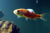
A Gold Fish with Fish Dropsy in containment. Notice the pinecone appearance
In dropsy, a concentration of fluid in the body tissues and cavities causes the fish's abdomen to become swollen and appear bloated (Ascites). Swollen areas may exhibit a 'pine-cone' appearance caused by the fish's scales sticking out. Fish may also stop feeding, appear off-color, become listless and/or lethargic, have sunken eyes, and hang at the top or stay at the bottom of the aquarium. The condition affects the fish's internal organs, ceasing proper function.
[citation needed]
Causes
The cause of fish dropsy can be difficult to diagnose. The main cause is bacterial infection. The causative agent may be introduced through poor water quality. Kidney failure or excess fluid (ascites) due to liver or heart failure are other possible causes.
[citation needed] Some household (pet) fish may produce a red film in their urine during pregnancy or change of habitat i.e.new fish tank relocation after only a weeks time.
[citation needed]
Treatment
Because dropsy is a symptom of an illness, it may or may not be contagious. However, it is standard practice to quarantine sick fish to prevent stress among the other fish in the tank community. This extra stress may make the others vulnerable to dropsy or other forms of disease.
Prognosis
Most cases of dropsy are fatal. By the time the fish has swollen up enough that the scales begin to raise, the internal damage may be too extensive to repair.
[citation needed]
Prevention
Water quality is an important factor in prevention of fish disease. Water changes will dilute existing disease agents, and reduce stress on the tank occupants.
[citation needed]
(5)Fin Rot
 Fin rot
 is a symptom of disease or the actual disease in fish. This is a disease which is most often observed in aquaria and aquaculture, but can also occur in natural populations
[1]. Fin rot can be the result of a bacterial infection (
Pseudomonas fluorescens, which causes a ragged rotting of the fin), or as a fungal infection (which rots the fin more evenly and is more likely to produce a white 'edge'). Sometimes, both types of infection are seen together. Infection is commonly brought on by bad water conditions, injury, poor diet, or as a secondary infection in a fish which is already stressed by other disease. Fin rot starts at the edge of the fins, and destroys more and more tissue until it reaches the fin base. If it does reach the fin base, the fish will never be able to regenerate the lost tissue. At this point, the disease may attack the fish's body directly.
Very Common In:
- Betta fish; due to poor water conditions.
Symptoms:
- Fin edges turn white
- Fins fray
- Base of fins inflamed
- Entire fin may rot away or fall off in large chunks
Ways to treat:
- Change the water
- Treat with antibiotics like Melafix
- Use aquarium salt to keep water healthy
- Find out the ph and correct it
- Use Stress Coat
Prevention:
- Make sure the water is good quality
- Feed fresh food in small portions
- Maintain constant water temperature
(6)Trematoda:
Trematoda is a class within the phylum Platyhelminthes that contains two groups of parasitic flatworms, commonly referred to as "flukes".
 Taxonomy and biodiversity
The trematodes or flukes are estimated to include 18,000
[1] to 24,000
[2] species, and are divided into two subclasses. Nearly all trematodes are parasites of mollusks and vertebrates. The smaller Aspidogastrea, comprising about 100 species, are obligate parasites of mollusks and may also infect turtles and fish, including cartilaginous fish. The Digenea, which constitute the majority of trematode diversity, are obligate parasites of both mollusks and vertebrates, but rarely occur in cartilaginous fish. Formerly the Monogenea were included in Trematoda on the basis that these worms are also vermiform parasites, but modern phylogenetic studies have raised this group to the status of a sister class within the Platyhelminthes, along with the Cestoda.
Anatomy
Trematodes are flattened oval or worm-like animals, usually no more than a few centimetres in length, although species as small as 1 millimetre (0.039 in) and as large as 7 metres (23 ft) are known. Their most distinctive external feature is the presence of two suckers, one close to the mouth, and the other on the underside of the animal.
[3] The body surface of trematodes comprises a tough syncitial tegument, which helps protect against digestive enzymes in those species that inhabit the gut of larger animals. It is also the surface of gas exchange; there are no respiratory organs.
[3] The mouth is located at the forward end of the animal, and opens into a muscular, pumping pharynx. The pharynx connects, via a short oesophagus, to one or two blind-ending caeca, which occupy most of the length of the body. In some species, the caeca are themselves branched. As in other flatworms, there is no anus, and waste material must be egested through the mouth.
[3] Although the excretion of nitrogenous waste occurs mostly through the tegument, trematodes do possess an excretory system, which is instead mainly concerned with osmoregulation. This consists of two or more protonephridia, with those on each side of the body opening into a collecting duct. The two collecting ducts typically meet up at a single bladder, opening to the exterior through one or two pores near the posterior end of the animal.
[3] The brain consists of a pair of ganglia in the head region, from which two or three pairs of nerve cords run down the length of the body. The nerve cords running along the ventral surface are always the largest, while the dorsal cords are present only in the Aspidogastrea. Trematodes generally lack any specialised sense organs, although some ectoparasitic species do possess one or two pairs of simple ocelli.
[3]
Reproductive system
Most trematodes are simultaneous hermaphrodites, having both male and female organs. There are usually two testes, with sperm ducts that join together on the underside of the front half of the animal. This final part of the male system varies considerably in structure between species, but may include sperm storage sacs and accessory glands, in addition to the copulatory organ, which is either eversible, and termed a
cirrus, or non-eversible, and termed a penis.
[3] There is usually only a single ovary, which is connected, via a pair of ducts to a number of
vitelline glands on either side of the body, that produce yolk cells. Eggs pass from the ovary into a glandular receptacle called the
ootype or
Mehlis' gland, where fertilisation occurs. This opens into an elongated uterus that opens to the exterior close to the male opening. The ovary is often also associated with a storage sac for sperm, and a copulatory duct termed
Laurer's canal.
[3]
Life cycles
Main article: Trematode lifecycle stages
Almost all trematodes infect mollusks as the first host in the life cycle, and most have a complex life cycle involving other hosts. Most trematodes are monoecious and alternately reproduce sexually and asexually. The two main exceptions to this are the Aspidogastrea, which have no asexual reproduction, and the schistosomes, which are dioecious. In the definitive host, in which sexual reproduction occurs, eggs are commonly shed along with host feces. Eggs shed in water release free-swimming larval forms that are infective to the intermediate host, in which asexual reproduction occurs. A species that exemplifies the remarkable life history of the trematodes is the bird fluke,
Leucochloridium paradoxum. The definitive hosts, in which the parasite multiplies, are various woodland birds, while the hosts in which the parasite grows (intermediate host) are various species of snail. The adult parasite in the bird's gut produces eggs and these eventually end up on the ground in the bird's faeces. Some very fortunate eggs get swallowed by a snail and here they hatch into tiny, transparent larva (miracidium). These larvae grow and take on a sac-like appearance. This stage is known as the sporocyst and it forms a central body in the snail's digestive gland that extends into a brood sac in the snail's head, muscular foot and eye-stalks. It is in the central body of the sporocyst where the parasite replicates itself, producing lots of tiny embryos (redia). These embryos move to the brood sac and mature into cercaria.
Infections
Main article: Trematode infection
Human infections are most common in Asia, Africa, South America, or the Middle East. However, trematodes can be found anywhere that human waste is used as fertilizer.
Etymology
Trematodes are commonly referred to as
flukes. This term can be traced back to the Old English name for flounder, and refers to the flattened, rhomboidal shape of the worms. The flukes can be classified into two groups, on the basis of the system which they infect in the vertebrate host.
- Tissue flukes infect the bile ducts, lungs, or other biological tissues. This group includes the lung fluke, Paragonimus westermani, and the liver flukes, Clonorchis sinensis and Fasciola hepatica.
- Blood flukes inhabit the blood in some stages of their life cycle. Blood flukes include species of the genus Schistosoma.
They may also be classified according to the environment in which they are found.
(7)Velvet (fish disease)
 velvet disease
, also called
gold dust disease is a fish disease caused by the dinoflagellate parasites of the genera
Oödinium,
Amyloodinium or
Crepidoodinium which gives the fish a dusty, slimy look.
[1] The disease often infects fishes in tropical aquaria. The parasite is single-celled and enters the slime coating of a host fish in its motile juvenile stage where it matures. The mature parasites break through the slime layer and drop to the bottom of the aquarium and attach themselves to solid surfaces. Here they form a cyst, which develops into numerous new juvenile individuals known as
tomites. Velvet is highly contagious and can prove fatal to fish. The infected fish usually swims around scratching at objects in the tank very rapidly and usually has its fins very close to its body.
(8)Ammonia poisoning
Ammonia poisoning is a common fish disease in new aquariums, especially when immediately stocked to full capacity. Ideally, the level of ammonia (NH
3) and ammonium compounds (i.e. those containing NH
4+) should be zero. Although trace amounts are generally harmless, they can still lead to problems over time. Understanding the nitrogen cycle is essential for the keeping of any aquatic life. The amount of ammonia present is usually accompanied by a rise in pH. As ammonia is a base, it is stabilized by alkaline water. It can cause damage to the gills at a level as small as 0.25 mg/L.
Diagnosis
A history of the tank: filter changes, power outages, excessive feeding, or the addition of microbicidal or antibiotic agents to aquarium can aid in diagnosis. An ammonia test is the most sure way of diagnosing ammonia poisoning.
Symptoms include:
- Purple, red or bleeding gills
- Fish may clamp, may appear darker in color
- Red streaking on the fins or body
- Fish may gasp for air at the surface of the tank water
- Torn & jagged fins
- Fish may appear weak and lay at the bottom of the tank
Prevention
The nitrogen cycle in an aquarium.
Ammonia poisoning is impossible to cure, however it can be prevented easily by first cycling the tank (see below). Treatments include immediately reducing the ammonia level through many small water changes. Alternatively an ammonia detoxifier can be used, though such chemicals are best used in emergencies only, and do not provide a substitute for adequate tank cycling. Once the ammonia is removed, the fish may recover if the damage is not too extensive. Increasing aeration may be desirable, as the fishes' gills are often damaged by the ammonia. This can increase the probability of survival slightly. Also, all other sources of stress should be removed, and the cause of the ammonia should be addressed.
Prevention (Tank Cycling)
Tank cycling is a process during which ammonia reducing bacteria are built up sufficiently to handle the tank load. This process can take two to four weeks.
 Lernaea (also spelled Lernea) is a genus of copepod crustaceans commonly called anchor worms, parasitic on freshwater fishes. They mate during the last free-swimming (copepodid) stage of development. After mating, the female burrows into the flesh of a fish and transforms into an unsegmented, wormlike form, usually with a portion hanging from the fish's body.[1]


good post ......
ReplyDelete